


Osteoma is a degenerative disease of the joints and discs. Osteoarthritis affects the cartilage tissue of the discs. As a result, the discs harden and contract, losing their cushioning properties and causing severe pain.
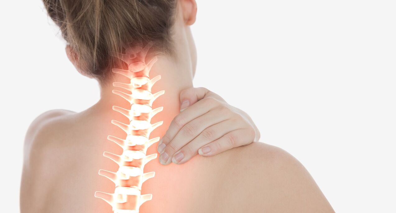
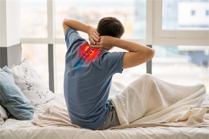
Cervical osteosarcoma is a progressive degenerative-dystrophic process that leads to deterioration, deformity and destruction of the intervertebral discs of the cervical region. The loss of shock-absorbing cartilage causes pain both from contact of the joint surface (spondylolisthesis) and from compression of the nerve roots of the spinal cord.
If not treated in time, the spine can degenerate due to loss of natural flexibility, impaired blood supply to the brain, impaired nerve conduction in parts of the body to conduct internal conduction. roots of the cervical spine.
Pathology can develop independently and as part of a generalized spinal cord injury, including the thoracic, lumbar, and sacral regions.
General information about cervical osteonecrosis
It is believed that osteonecrosis of the cervical spine is more common than other parts. In fact this is not the case - dystrophy develops evenly at all points of maximum load - in the area \u200b \u200 main flexors of the spine (the lower the part lies, the greater the load will be). great) . However, the symptoms of cervical osteonecrosis are more pronounced, so they seem to occur more often. This is due to the high mobility of the cervical vertebrae, which simultaneously hold the head, as well as the special position of the outlet of the roots of the spinal cord.
On a note!According to statistics, the disease affects more than 60% of middle-aged and older people. However, recently there has been a rejuvenation of the pathological process - pathology occurs in young people and even in adolescents. This is due to the general computerization of learning and working, as well as a decrease in physical activity and a decline in nutritional quality.
Taking into account the age subject, it is possible to distinguish 2 forms of cervical osteonecrosis - physiological and pathological.
physiological processAssociated with the natural aging process of the body, when the symptoms of the disease are the result of the discs being worn down gradually. The process occurs under the influence of the endocrine system and is the result of changes during menopause. The destruction of cartilage structures begins at the center of the disc and is accompanied by the gradual replacement of cartilage tissue by fibrous tissue. This pathology is irreversible, but can be compensated with special drugs.
Pathological processassociated with abnormal destructive changes in the body - immunity, dystrophy, inflammation, metabolism. First of all, related to cartilage tissue - salt deposition appears on bone structures, nerve roots become inflamed, atrophy or increase skeletal muscle tone, leading to impaired blood circulation in the head and neck region- chest. With timely diagnosis, the pathology is treatable and ends with the complete restoration of healthy functioning of organs and tissues.
Stages of cervical osteonecrosis and their symptoms
There are 4 main stages of the pathological process:
- Stage 1 - manifested by slight discomfort and muscle tension in the diseased area, instability of the cartilage discs;
- Stage 2 - there is local pain, especially when moving the head. The discs are deformed, the capsule begins to collapse, the distance between the vertebrae decreases;
- Stage 3 - pain gradually increases and becomes constant, movement is limited. Turning the head can cause dizziness, nausea, reduced blood supply to the brain leading to generalized coma, fatigue, impaired concentration, cartilage becomes thinner, vertebrae close, capsular capsule becomes damaged. complete destruction with the risk of disc herniation;
- Stage 4 - pain syndrome that completely immobilizes the neck region; blood circulation of the brain is impaired and requires constant medical support; The vertebrae begin to grow together.
Cervical fibroids: signs, symptoms, pathological treatment

In the early stages, osteonecrosis is asymptomatic. As the disease progresses, a prominent feature is the presence of painful or uncomfortable sensations in the head, neck and chest, less often in the upper extremities.
All possible symptoms can be attributed to 4 types of syndromes: cardiac, vertebral, lens (nerve) and vertebral artery syndrome (with circulatory disorders).
Vertebral syndrome:
- crunching in the neck when turning / tilting the head;
- as the disease progresses, pain and difficulty in movement occur;
- Structural morphological disorders in the vertebral body and disc space (visible on radiographs).
Heart syndrome:
- shortness of breath, weakness;
- feeling of incomplete inspiration, lack of atmosphere;
- spontaneous phenomena from the cardiovascular system - angina pectoris, post-vascular pain, burning;
lens syndrome:
- numbness of the tongue, shoulders, fingers, occipital region;
- difficulty swallowing;
- discomfort in the area between the shoulder blades;
- occipital and forehead headache.
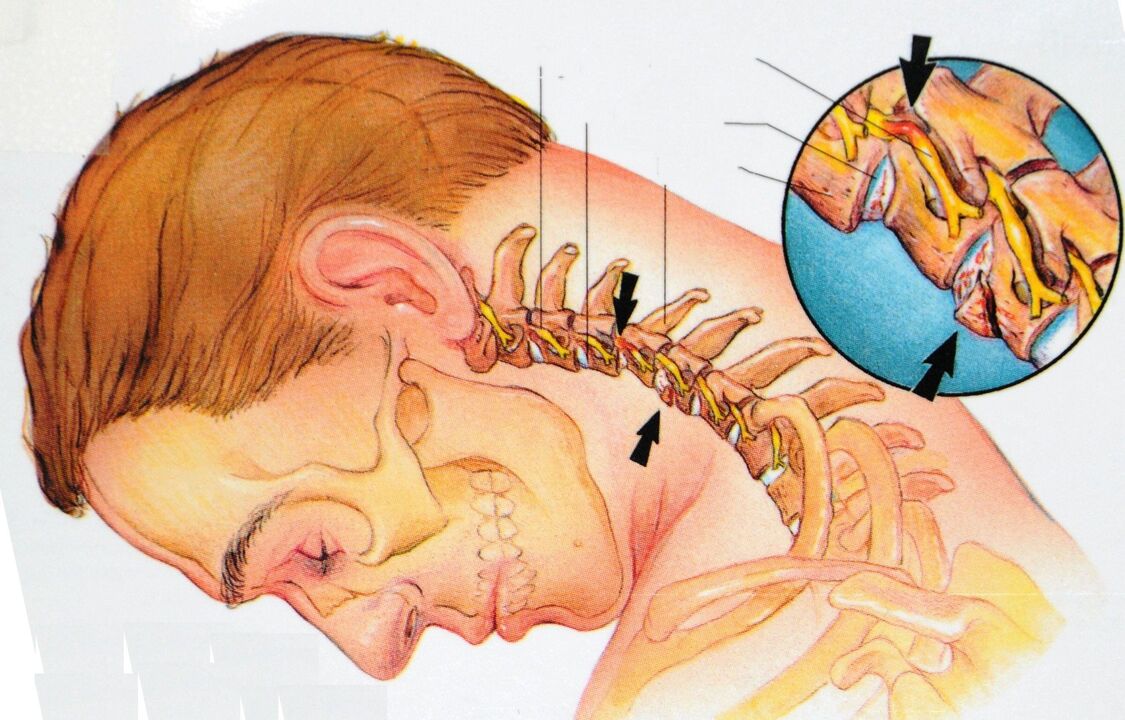
vertebral artery syndrome:
- unreasonably high blood pressure;
- dizziness, up to loss of consciousness;
- tinnitus, feeling of having cotton balls in the head;
- temporary unilateral blindness, "flies" in the eye;
- cyclical nausea, especially when moving the head;
- headaches - mainly in the back of the head, as well as migraines;
- drowsiness, decreased performance, memory, concentration, depression.
Attention!All of these syndromes must be combined. The absence of symptoms of one of them may be the reason for the differential diagnosis from other groups of diseases.
Causes of cervical osteonecrosis
Cervical dystrophy is related to the vertical position of the skeleton and the specific distribution of static and dynamic loads, largely dependent on the prevailing posture and the degree of musculoskeletal development. .
- lack of movement - what does not grow - deterioration: muscles weaken, tissues are destroyed;
- incorrect static posture - muscle clamping leads to circulatory disorders with subsequent tissue degeneration;
- lack of nutrition or unbalanced diet - the body must receive everything necessary for the building and renewal of the bone and cartilage structure of the skeleton, maintaining muscle tone;
- obesity, overweight, carrying heavy loads - the load on the skeletal structure increases;
- constant nervous tension and nervous tension;
- hypothermia of the cervical region - "cold", "bulging" - provokes hidden inflammatory processes;
- the presence of autoimmune diseases associated with cartilage that leads to its premature destruction;
- endocrine diseases that disrupt mineral metabolism, reduce absorption of calcium, silicon, phosphorus and other elements of bone and cartilage tissue;
- neck injury;
- Congenital malformations of the spine and adjacent muscles.
Diagnosis of cervical osteonecrosis
The diagnosis of "cervical spondylosis" is difficult due to the low specificity of the symptoms and their variety of manifestations. During the examination, you will need to consult a neurologist, surgeon, orthopedist, cardiologist.
Physical examination is performed by a doctor with a patient question. The primary diagnostic load lies on instrumental and laboratory research methods.
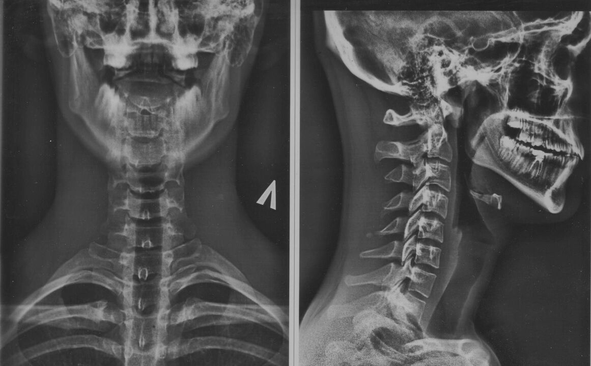
- X-ray of the cervical region; At an early stage in the process, an MRI of the cervix will be more informative - it will provide high quality images of the hard and soft tissues - it will show the condition of the discs, the presence ofof bone cells, malformations, damage to nerve roots and blood vessels; assessment of the condition of ligaments, muscles, bone tissue; shows the dynamic state of soft tissues;
- dopplerography of the cervical vasculature will help to assess the hemodynamics and extent of vascular damage (specifically, the state of the vertebral arteries);
- contrast myelogram - will help to deal with nerve processes that are suspected of being intrusive;
- Electrocardiography and echocardiography are used in the differential diagnosis of cardiac syndromes from cardiovascular diseases.
How to treat cervical osteonecrosis?
A complex of therapeutic measures is formed taking into account the stage of the disease (acute, chronic), the degree of damage and the etiology of the pathology. Use conservative treatment, surgery, a mixed approach.
conservative effect
This is a gradual recovery or compensatory damage against the background of symptomatic treatment. Includes drug therapy, physical therapy, exercise therapy, and massage methods.
Medical treatment:
- pain relievers - mainly gels and ointments applied to the skin; in severe cases - general pain relievers in the form of tablets;
- anti-inflammatory drugs - NSAIDs, as well as corticosteroids (short course if necessary);
- drugs to improve microcirculation and general blood circulation;
- chondroprotectors - means protection and restoration of cartilage tissue;
- muscle relaxants - to eliminate clamping and muscle spasms;
- complex of vitamins and microelements - necessary for nutrition and supporting tissues with building factors.
As the acute symptoms subside, the methods of physical therapy, exercise and self-massage are connected.
Physical therapyImproves nutrition of cartilage and bone tissues by restoring blood supply in the damaged area. In order to avoid complications, the method of isostatic motion should be used, when instead of the actual head rotation and potentially injurious head tilt, their imitation is used.
Attention!All actions should be taken only after diagnosis and consultation with a doctor.
This technique allows you to develop and strengthen the atrophied short muscles of the neck and stabilize the position of the cervical spine. Sequence of basic exercises:
- Place your right palm on the side of your head - for 10 seconds, press your palm to your head, while contracting the muscles of your head and neck to create resistance - your head should be motionless.
- Lower your arms, relaxing your head and neck muscles as much as you can for 20 seconds.
- Repeat the exercise with your left hand.
- Place your hands on your forehead with your palms - for 10 seconds, press on your forehead as if trying to tilt your head back, while contracting your neck muscles to counteract the movement. The head must be motionless.
- Lower your arms, relaxing your muscles as much as you can, similar to the previous exercise.
- Place both palms in the upper area behind the head. In the same way, apply pressure to the neck muscles, trying to tilt the head forward - the head should remain motionless.
- Lower your arms, relaxing the muscles in your neck and head. Repeat the movement 4-10 times.
After strengthening the short muscles of the neck, you can begin to perform dynamic exercises.
On a note!Swimming and water gymnastics have been shown to be a technique for restoring cervical mobility.
Self-massageallows you to reduce the intensity of local reactions and reduce muscle clamping forces during static work. Enforcement rules:
- zone of influence - the back of the head, the back and side of the neck;
- perform the procedure in a sitting position;
- movements should be performed in the direction from the spine;
- use only fingertips;
- avoid pressure on inflamed areas;
- Make movements smooth - sharp pressure can be harmful.
Physical therapyTypical for inpatient treatment and spa rehabilitation. Proven:
- electrophoresis - warms the area, improves microcirculation, is used for deeper penetration of topical preparations;
- acupuncture therapy;
- amplipulse;
- UHF.
Surgical intervention is indicated for complex extrusion syndromes, spinal cord invasion and intractable pain syndromes.
What are dangerous cervical fibroids?
The neck region concentrates a dense interlacing of major blood vessels, neural processes and the dynamic structure of the skeleton. In the absence of treatment, serious pathological changes can be observed:
- weakening of the annulus causes dislocations and dislocations in the region of the most mobile vertebrae;
- the presence of osteogenic substances and muscle spasms lead to invasion of nerve roots and blood vessels with the formation of compression syndromes;
- destruction of the cartilaginous discs and the convergence of the vertebrae leads to disc herniation with invasion of nerve tissue.
Each of these phenomena is followed by a markedly negative reaction from the organism as a whole.
Possible complications and consequences
The list of the most common complications of cervical osteonecrosis includes:
- vegetative dystonia;
- hypertension;
- oxygen starvation of the brain and its structures;
- retinal dystrophy, visual impairment;
- malfunction of the thyroid gland;
- dysfunction of the esophagus and trachea - difficulty swallowing and spasms of the airways;
- intractable pain in the head, neck, chest, upper extremities;
- convulsions and numbness of the face and hands;
- Disruption of the pituitary-hypothalamic system, which entails the failure of the entire hormonal activity of the body.
Measures to prevent cervical osteonecrosis
The most effective treatment is prevention. Prevention will help you with this. Just following a few basic recommendations should suffice:
- correct your posture,
- create a comfortable workplace;
- during sedentary work, take a break for a "minute of physical education";
- include in your diet foods rich in calcium, magnesium, phosphorus, silicon - fish, nuts, seeds, legumes, dairy products, fresh vegetables, fruits; limit eating salty, sweet, starchy, hot and spicy foods;
- for sleep and rest use orthopedic mattresses and pillows;
- take up a sport that doesn't involve endurance - you'd better prioritize swimming.
Even if you cannot take into account all the requirements, moderate exercise, proper nutrition and attention to your posture can significantly reduce the risk of developing the pathology.
The potential cause of impotence and male and female infertility is osteonecrosis.
Even in school, in biology classes, they tried to convey important information about the great role of the spine in maintaining human health. Unfortunately, many people then got involved in more important things and didn't listen to the teacher's words. But to no avail! Doctors say that disorders of the musculoskeletal system, especially due to osteonecrosis, can provoke the development of a number of different serious diseases.
Why does osteonecrosis have a strong destructive effect on the human body
Often men suffer from impotence and infertility, while women try in vain to get pregnant and do not even suspect that this may be caused by normal osteonecrosis. The fact is that the cause of the development of the disease lies in the violation of the blood supply to the tissues of the vertebrae and the muscles that surround them. Since there are no blood vessels in the intervertebral disc, they are the first to suffer from a lack of water and other substances. This leads to cartilage cracking, which means that the discs are no longer able to absorb the load on the spine. This is the cause of chronic back pain.
If you do not intervene at this stage, the process of bone necrosis will continue to progress and cause complications, such as a herniated disc. This, in turn, causes a violation of the segmental apparatus of the spinal cord and impairs blood circulation, including in the pelvic organs. This is the main reason for the development of many disorders in the work of internal organs, as well as impotence and infertility that concern us.
The development of impotence in men, in addition to violations at the physical level, also contributes to psychological factors. After all, for every normal, full-fledged man, even a single failure in bed becomes a drama and it doesn't matter if it's due to an exacerbation of osteonecrosis, nerve pain. back pain or other causes of back pain.
Men may begin to have difficulty getting an erection when they have degenerative disc disease in the cervical or lumbar spine. But in each case, the pathology develops according to its own mechanism.
Cervical bone necrosis
When suffering from this type of disease, it will reduce the quality of blood circulation to the brain, causing problems in the production of sex hormones and substances responsible for blood vessel tone. Therefore, with cervicitis, most often patients complain of decreased libido, lack of qi, and problems with ejaculation.
Lumbar bone necrosis
Since the pelvic region, specifically the male genitalia, is in an "off" state due to a spinal cord disorder, nerve impulses are not always able to travel to the genitals, resulting in dysmenorrhea. erectile dysfunction.
Male and female infertility as a result of osteonecrosis
Often, in the absence of other prerequisites, unsuccessful attempts at conception in both men and women can be the result of pathological processes that accompany osteonecrosis. Most often, the cause of the problem lies not in a violation of the blood supply to the organs located in the small pelvis, but in neurological disorders.
Examination of infertile women revealed degenerative changes in the lower thoracic spine and lower back. In infertile men, osteonecrosis affects the radial region. Such differences in the areas affected by osteonecrosis are explained by the structural and visceral specificity of the pelvic organs in representatives of different sexes.
Sometimes women can't get pregnant even if they don't experience any signs of the disease and don't feel even the slightest discomfort in their backs. This is mainly due to the fact that disorders in the reproductive system can occur even if only the anterior roots of the spinal cord are injured, without pain.
Therefore, everyone with sexual and reproductive dysfunction is recommended to have the most comprehensive examination, not forgetting a neurologist and a chiropractor. It is possible that the root of the problem lies precisely in the pathologies of the spine.