


Arthrosis of knee joints is a chronic (long -term) degenerative disease that causes the destruction of cartilage in the joint.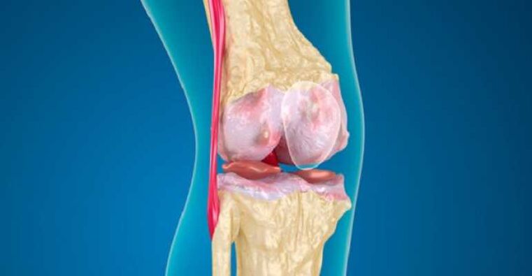 Symptoms include pain, hardness and swelling.Treatment options to reduce pain and defects include lifestyle changes (diet, physical exercises), treatments, drugs and surgery.
Knee fibrosis
Osteoarthritis is a common disease, accompanied by chronic pain, fatigue.Recent clinical data shows that the sensitivity of the center stimulates the deformed osteoarthritis of the knee joint.An understanding of improvement in how knee arthritis affects the central pain treatment is very important to determine new pain relief targets/new treatment strategies.
The Cannabinoid receptors inhibit the function of peripheral immune cells and adjust the central neurological immune answers in neurological degenerative models.The whole body birth of the receptor owner weakens the pain caused by OA, and the changes in the circulation of anti -inflammatory and anti -inflammatory cytokines have been manifested in this model.
Deforming joint diseases
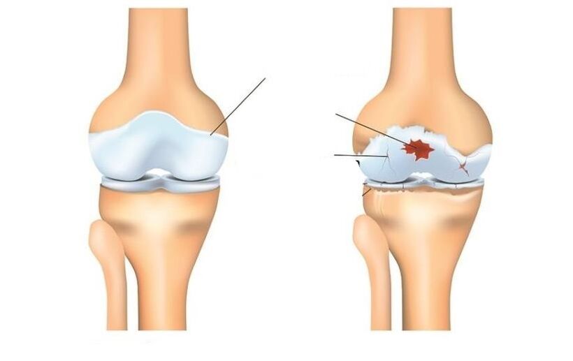
The deformation of the knee joint is inflammation and wearing cartilage on the bone forming the knee joint (osteo = bone, artro = joints, itis = inflammation).Diagnosis of osteoarthritis of the knee joint is based on two main results: X -ray data on changes in bone health (using medical images, such as X -rays and magnetic resonance images of MRI) and human symptoms.About 14 million people have symptoms of knee arthritis.Although the elderly became more popular, 2 million out of 14 million people with knee arthritis symptoms were under 45 years old while diagnosis, and more than half a younger than 65 years old.
Osteoarthritis (OA's knee) is a disease that is caused by inflammation and osteoarthritis, eventually worse.
This affects the entire joint, including bones, cartilage, ligaments and muscles.Its progress is affected by age, body mass index (BMI), bone structure, geneticist, muscle strength and activity level.OA knee can also grow as a secondary state after knee injury.Depending on the stage of the disease and the presence of trauma or the condition related to it, the knee may be controlled by physical therapy.Serious or more expansion cases may require surgical intervention.
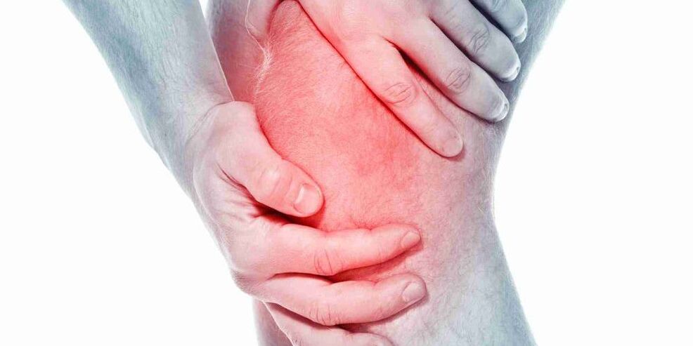
Symptom
Developers of arthritis can undergo a variety of symptoms and restrictions based on the progression of the disease.The pain occurs when the cartilage covers the knee bone and knee.The areas where cartilage is worn or damaged, exposing the lower bones.The effect of bone allows you to increase stress and compress cartilage, and sometimes exposure to the bone when moving, can cause pain.Because the knee is a joint, the level of activity, the extent of the activity, as well as the type and action time, as a rule, has a direct impact on symptoms.Symptoms may worsen with weight activity, for example, when walking with a heavy object.
The symptoms of knee joints may include:
- Relieve pain during or after surgery, especially when walking, climbing, lowering the stairs or moving from a seating position to a standing position.
- Pain or hardness after sitting with knees or knees straight for a long time.Pain is the most common symptom of osteoarthritis.As the disease progresses and inflammation progresses, the pain may become unchanged.
- A feeling of jumping, cracking or grinding when moving the knee.
- Swelling after action.
- The hardness of the affected joint is usually seen first in the morning and after rest.
- Edema, sometimes warm when touching, may notice in arthritis joints.
- Deformation can occur with osteoarthritis due to bone growth and cartilage loss.The growth of the bone in the end joints of the finger is called the Hyberden buttons.The bushar buttons are the development of the bone in the middle joints of the finger.The degeneration of the cartilage of the knee joint can lead to the external curvature of the knee (foot onions).
- A cracked sound or a mesh feeling may be noticed when arthritis moves.This is caused by wiping bones against bones or raw cartilage.
Usually these symptoms do not arise suddenly and all at the same time, but gradually develop over time.Sometimes people do not admit that they have osteoarthritis, because they cannot recall a certain time or injury causing their symptoms.If the knee pain has worsened for a few months, this does not respond to resting or changing in activity, better than seeking advice for a medical staff.
Diagnose
Osteoarthritis can usually be diagnosed by the characteristic symptoms of pain, relieving motion and/or deformity.Osteoarthritis can be confirmed by X-ray or MRI scanning.General data includes narrowing the joint space between bones, loss of cartilage and spearhead or bone growth.Blood tests may be used to eliminate other possible conditions, but they cannot diagnose osteoarthritis.
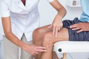
In knee arthritis, 2 main processes are diagnosed.First based on a report on symptoms and clinical examination.Physiotherapists will ask questions about history and health activities.The therapist will conduct physical examination to measure the movement of the knee (the range of motion), the power, the ability to move and flexibility.They may also require various movements to view, increase or relieve pain.
The second tool used to diagnose knee joints is a diagnostic image.Physiotherapists can send to the doctor, who will prescribe the X of the knee in different positions to check the damage to the bones and cartilage of the knee joint.
If the damage is suspected of being more serious for joints, you can order MRI to study the general condition of joints and surrounding fabrics.
Blood tests may also be required to eliminate other conditions that can cause symptoms similar to osteoarthritis of the knee joint.
Treatment
Depending on the severity of arthritis and the age of the patient, it will be chosen to treat the joints of the knee joint.Treatment may include operating methods or inactive, or their combination.
The first lines of arthritis of the knee joint include modification of activity, anti -inflammatory and weight loss drugs.Out of action to enhance pain can make this condition accepted by some people.Anti-inflammatory drugs, like iBuprofen and newer COX-2 inhibitors, helps reduce inflammation that can contribute to pain.
Physical therapy to strengthen the muscles around the knee can help absorb a part of shock given to the joint.This is especially true for arthritis of the knee cup (Patello-Femoral).Special types of braces are designed to convey to part of the knee joint, less than arthritis, which can also reduce pain.Injecting drugs inside the knee joint can also help temporarily.
In addition, walking with a stick in the hand on the opposite side, because a pain knee can help distribute a part of the load, relieve pain.Finally, weight loss helps reduce the force through the knee joint.The combination of these non -active measures can help relieve pain and defects caused by arthritis of the knee joint.
If the inactive methods do not allow you to withstand, the operation may be the best option for treating arthritis of the knee joint.The type of operation accurately depends on age, surgery and the main state.Some examples of surgical options to treat arthritis include bone surgery, including bone cutting for joint alignment and knee replacement surgery.
Modern methods to treat knee joints include bone surgery, this is a good choice if a young patient and arthritis are limited by a knee area.This allows the surgeon to rebuild the knee to remove the arthritis area and perform relatively non -load loads related to the parts of the knee joint.For example, the patient may be rebuilt to redistribute the load through the joints.The advantage of this type of surgery is that the patient's knee joint is preserved and has the ability to ensure many years of pain relief without fake knees.Disadvantages include a longer rehabilitation course and the ability to develop arthritis in the knee recently balanced.
Activities to replace knee joints include cutting joints and inserting fake joints.All joint surfaces are replaced, including the femur, lower leg and knee cup.The joint surfaces are removed, and the heads of the bone are replaced by a prosthetic leg.The prosthetic composition is usually made from metal and plastic surfaces, designed to slide smoothly together.
Replace knee joints
Overall activity to replace knee joints was first performed in 1968 and over the years developed in a reliable and effective way to get rid of the pain when turned off and allow patients to continue their positive life.Improvements in the field of surgery and transplant have helped turn this into one of the most successful orthopedic processes today.As the population becomes older and still more active, the demand for common knee replacement continues to increase.Many activities to replace knee joints took place in special surgical hospitals.Improvements in surgical technology and transplant design are some contributions that surgeons make.
People often wonder when and why they should replace their knees.This is a personal question depending on the extent of human activity and functional needs.Many people suffer from arthritis with pain, preventing them from participating in activities;Others are so weak that it is difficult to wear shoes and socks.A complete replacement of the knee joint provides solutions for muscle disease and is done to relieve pain and continue to work.After rehabilitation from the complete replacement of the knee joint, the patient may expect surgery, without pain.A complete replacement of the knee joint helps significantly improve the patient's condition and significantly reduces the cost of long -term treatment.This research shows that not only the overall replacement of the knee joint is economically effective compared to non -surgical control, but also provides larger function and the best quality of life.
A complete replacement of the knee joint is considered a major activity and the solution is not trivial.Usually people decided to undergo a surgery when they felt that they could no longer live with arthritis.Implant consists of 4 parts: tibia, femur, plastic and tea bone.The components of the tibia and the femur are made of metal, usually chromium, used to close the ends of the thigh and lower leg after removing the joints.Plastic insertion is made of polyethylene of extremely high molecular weight and fits the tibia bone component, so the thigh surface is polished along the plastic.The composition of the knee cup also glides on the front of the femur.Usually they are attached to bone cement.
The full replacement of the knee is performed in the operating room with a special Laminar airflow system, helping to reduce the possibility of infection.Your surgeon will wear a Viking space outfit, also designed to reduce the possibility of infection.The entire surgical group will include your surgeon, from two to three assistants and nanny.
Anesthesia is given through an outer catheter, a small tube that is put into the back.This is the same type of anesthesia for women when giving birth.During the surgery, the patient may be alert and sleepy.
After introducing the epidural mass around your thigh, a tourniquet or a cuff will be placed.Horizontal bar will be exaggerated during operation to reduce blood loss.The reduction for the complete replacement of the knee is made along the front knee.The incision will be measured from 4 to 10 inches depending on the surgery.
The joint surface of the femur, lower legs and patella bones are exposed and removed by strength instruments.At the same time, the deformation of the knee is repaired and after surgery, the knee becomes more straight.The bone is ready to take an artificial knee joint, and then a prosthetic leg is inserted.During the closing process, two drainage systems are installed around the working area to help reless.Sapers are used to close the skin.
The whole activity will take from 1 to 2 hours.After that, the patient will be taken to the recovery room where the tests will be tested.Most patients may be taken to a regular room for a few hours;Others will have to stay in the hall to recover, according to the definition of the surgeon and anesthetist.
Patients often stay in the hospital for 3-4 days after adequate surgery to replace the knee.
Risks during surgery
Some risks of surgical procedures include blood loss, blood clot formation in the leg and probability of infection.The general ratio of these risks is very small.They should be discussed with the surgeon before starting the surgery.
Some risks of the presence of fake knees include the possibility of parts that may weaken or wear out over time, or fake parts that may be infected.Once again, these issues will be discussed with the surgeon.
After surgery after surgery
Immediately after complete surgery to replace the knee joint, the patient will fall into the recovery room.Most patients can go to a regular ward after a few hours, when the feeling of returning to the leg.A painful pump related to an epidural external catheter will be given, which will allow you to control when curing the pain.Most people are quite comfortable with the help of a painful pump.
On the day of surgery, you can do some exercises, as directed by physical therapist, including quadrilateral reduction and moving your legs up and down.Depending on the hobby of the surgeon, you can start bending your new knee right after the surgery or on its first day.Patients will be allowed to drink water after surgery to wet their mouths, but drink liquid or you can cause nausea.Patients will have a catheter in the bladder, so you don't need to worry about urinating.As soon as the foot is restored, it will be allowed to sit down, stand up and perform a few steps with a pedestrian and therapist.
The first day after operation will operate, developed to help become more moving.Pacrant will meet physical therapists who will guide additional exercises.In addition, they will help stand on their feet and take a few steps when walking.As a rule, patients will be allowed to drink pure liquid.
In the next few days, it will be easier and easier to move.The patient will be released from pain and catheter.Treatment of pain will be given in the form of tablets.On the second day after surgery, if the recovery signs are found in the intestine, it will be allowed to eat regular food.
Depending on your age, physical condition before surgery and insurance coating, patients may be a candidate for short -term accommodation in a rehabilitation organization.If not, the patient will be discharged home, and the physiotherapist will come to his house to continue rehabilitation.The coordinator will discuss these options with the patient and will help him plan to return home.
A return for activity will be guided by a surgeon and therapist.As a rule, patients can walk as much as they want 6 weeks after surgery.Patients can continue moving after 6 weeks.After 8 weeks, patients can continue the game in golf and swim;At 12 weeks they can play tennis.The surgeon will help decide which actions can be resumed.
What is physiotherapist
All physical therapists are prepared through education and clinical experience to treat different conditions or injuries:
- A physical therapist has experience in treating people with knee osteoarthritis and after surgery to replace knee joints.Some physical therapists have a practice with an orthopedic center.
- A physiotherapist is a certified orthopedic clinical expert.This physical therapist will have advanced knowledge, experience and skills that can be applied to a state.
- You can find physical therapists who have this and other accounting data with MRI, an online tool to help find physical therapists with specific clinical knowledge.
General advice when you can find a physiotherapist (or any other medical service provider):
- Receive recommendations from family and friends or from other medical service providers;
- Moving to the clinic for physical therapy to be hospitalized, you need to ask about the experience of physiotherapists in supporting people with arthritis.
During the first visit to the physiotherapist, you need to prepare to describe the symptoms as much as possible and report on the activities that worsen symptoms.