


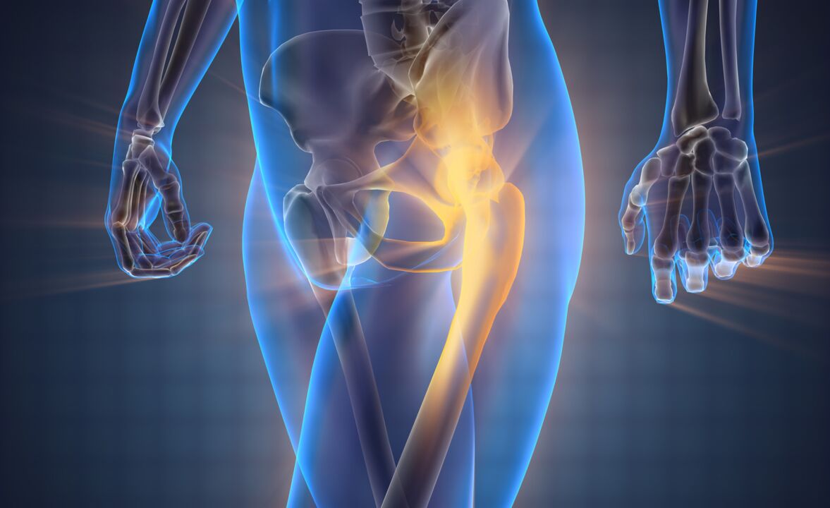
The hip joint, the largest joint in the human body, is subjected to daily stress from physical activity, supporting the body's weight. Many people think that joints only hurt when they get old. Of course, as we age, the layer of cartilage that absorbs shock when the joint is bent becomes thinner and the amount of fluid inside the joint decreases, leading to pain. However, not only age but also certain diseases contribute to pain that varies in intensity from mild to unbearable. The nature of pain in the hip joint can be dull, sharp, pressing or aching. Often depends on load, time of day and other factors. The cause of pain is determined by X-ray, CT, MRI, ultrasound, arthroscopy and other studies. Until the diagnosis is made, pain medication should be used and the rest of the lower extremities should be used.
Causes of pain in the hip joint
Soft tissue injury
The most common cause of acute pain is a bruise in the hip joint, caused by a fall to the side or by a direct blow, slightly limiting movement. Swelling may occur.
The pain syndrome gradually fades and disappears after a week. Damage to the ligaments in the hip joint often occurs due to traffic accidents and sports injuries, accompanied by a sharp pain syndrome accompanied by a feeling of cracking. The pain from swelling often increases again, moving down to the groin and thighs.
In case of ligament injury, the motor function of the lower limb is severely limited to the point of being unable to stand and, depending on the severity of the injury, such as: sprain, tear, rupture. The pain increases when the body leans in the opposite direction of the damaged ligament.
Bone and joint injuries
Femoral neck fractures often occur in older people due to trauma. A characteristic feature of osteoporosis is the presence of mild swelling without severe pain at rest. The pain increases strongly when moving. The symptom of a stuck heel is a typical sign when you cannot lift your leg straight up while lying down.
Due to high-energy trauma, young and middle-aged people often suffer pertrochanteric fractures, accompanied by sharp and deep pain. Movement is limited, unable to stand on lower limbs due to severe swelling of the joints.
Isolated fractures of the greater trochanter are rarely found in children and adolescents due to falls, direct impact, forceful muscle contractions, and are accompanied by acute, severe, extra-articular pain. . In this regard, patients avoid active movements.
The appearance of hip dislocation with acute unbearable pain occurs before falls from height, industrial and road injuries.
The leg may be bent or stretched due to joint deformity. When trying to stand on your feet or perform movements, an elastic gait appears, against the background of which there is severe pain, which does not subside until the joint subsides. Acetabular fractures develop independently or may result from hip dislocation. They are characterized by acute flare-ups of pain deep in the hip joint, making any movement difficult. The legs may become shortened and turned outward, making support impossible.
Degenerative process
In the early stages of coxarthrosis, after heavy exertion or at the end of the day, the patient begins to limp due to the appearance of dull, periodic pain that spreads to the hip or knee joints accompanied by slightly stiff movements. Increasingly, pain is noted not only during movement, but also at rest.
With severe coxarthrosis, the patient must rely on a cane. Movement is limited, the affected leg is shortened, this leads to increased load on the joint. The pain increases not only when walking but also when standing. Hip chondromatosis occurs like subacute arthritis. Moderate, transient pain accompanied by a crunching feeling and limited movement. When the nerve endings inside the joint are compressed, severe pain occurs, limiting movement. With hip arthritis, trochanteritis often develops, accompanied by inflammatory and degenerative damage to the tendons of the gluteus medius in the area of attachment to the greater trochanter. Pain syndrome appears when lying on the painful side, the pain increases when trying to move the hip to the side.
Bone nutrition problems
In children and adolescents, dull, deep pain in the knees and hips develops against the background of Perthes disease, which is characterized by necrosis of the femoral head. The pain increases after a few months, becoming constant, acute and debilitating. There is joint swelling, limited mobility and limping. After that, the pain syndrome gradually subsides and motor functions are restored in various ways.
Aseptic necrosis of the femoral head in adults occurs due to circulatory disorders and progresses similarly to Perthes disease, but is less favorable because half of the cases are bilateral.
At first, persistent pain occurs cyclically, then gradually increases, to the point where the patient loses the ability to completely stand on the leg due to joint destruction due to insufficient blood circulation. Gradually the pain syndrome subsides. Progressive limitation of movement over two years resulted from hip arthritis and lower limb shortening.
In the proximal metaphysis of the femur in boys aged 10-15 years, solitary bone cysts form, accompanied by mild periodic pain in the hip joint. In young children, there is no swelling. Because the symptoms do not appear, the reason to see a doctor is due to pathological bone fractures or increasing limitation of movement.
Hip pain may be due to avascular necrosis of the femoral head. The disease occurs due to circulatory disorders in the joints associated with long-term use of glucocorticoid hormones (they are prescribed for bronchial asthma, rheumatoid arthritis and some other diseases), alcoholism and severe diabetes. Joint necrosis may precede injury, but in some cases the actual cause cannot be determined. The pain in this case is very intense and occurs when walking and when trying to stand on the painful leg.
Arthritis
Wave-like pain ranging from mild to severe and continuous, with limited mobility in the hip joint in the morning, is a characteristic sign of aseptic arthritis. Symptoms such as stiffness, swelling, redness, increased body temperature, and pain when pressed will be noted.
Periodic pain in rheumatoid arthritis appears due to changes in weather conditions due to changing seasons, due to hormonal changes after childbirth or during menopause. The pain can be moderate and weak, persistent and aching, increasing sharply when palpated, accompanied by synovitis, edema, congestion, hyperthermia and limited movement.
Severe pain syndrome, jerking, tearing, both at rest and during movement, develops due to the spread of infection on the background of septic arthritis. Therefore, the genus has an obligatory position. The disease is accompanied by fever, chills, sweating, severe weakness, swelling, redness of the joints and increased temperature. If left untreated, bacterial septic arthritis can develop into panarthritis - severe purulent inflammation of the hip joint with acute sharp pain, severe fever, weakness, fainting, increased blood pressure andincreased body temperature.
Other inflammatory disorders
Against the background of open fractures, postoperative wounds, due to the appearance of pus, pain in the hip joint due to osteomyelitis increases for 1-2 weeks with signs of inflammation. Synovitis, tendinitis and bursitis develop with injuries and other diseases of the hip joint, and rarely become manifestations of allergies. In acute synovitis, the joint is slightly tender but the pain may increase due to swelling and increased fluid within the joint. Chronic synovitis with mild pain. With intermittent hydroarthrosis, the hip joint is slightly painful, accompanied by limited mobility, disappears within 3-5 days and recurs again after a certain period of time due to fluid accumulation in the joint.
Specific infections
With tuberculosis of the hip joint, weakness and fatigue will first appear, then a feeling of weak twitching or muscle pain when walking will appear. The patient begins to lose limbs. As the disease progresses, pain radiates down to the knee combined with swelling, redness, and synovitis. Pulling, twisting pain along with fever, lymphadenopathy, and skin rash may occur in acute brucellosis. During the chronic course of the disease, deformities form over time.
Congenital disease
Hip dysplasia is determined by the degree of incompatibility between the femoral head and the acetabulum. With congenital dislocation, pain appears as soon as the child begins to walk, accompanied by limping. With moderate subluxation, pain occurring at the age of 5-6 years is related to the load on the leg. With subluxation, pathology occurs for a long time without symptoms; with the development of dysplastic coxarthrosis at the age of 25-30, pain occurs at rest, intensifies with movement. All forms of dysplasia are accompanied by asymmetry of skin folds and limited mobility. In case of dislocation, the leg is shortened.
Tumor
The first painful symptoms of benign tumors are often mild and unstable, and do not progress over a long period of time. The growth of the tumor causes pain in the hip area to gradually increase. Malignant tumors (osteogenic sarcoma, chondrosarcoma) are characterized by mild, short-term pain, sometimes worse at night. After that, the pain symptoms become acute, continuous, cutting, enveloping, spreading to the entire joint, swelling and deformity. The patient lost weight, was weak and had a mild fever. In severe cases, the pain becomes so intense and unbearable that it can only be eliminated with the help of anesthetics.
Other reasons
Hip pain sometimes appears in the lower back, in the back due to neuropathy of the sciatic nerve, but gradually reduces compared to severe pain in the back of the buttocks and thighs, weakness of the lower limbs with sensory disturbances. . Dull and aching pain occurs with osteoarthritis, disc herniation, spondylitis, spinal deformity and spinal curvature due to joint overload, development of coxarthrosis and mental illness.
Diagnose
For initial diagnosis, the involvement of a general practitioner is required. Trauma diagnostic measures are performed by the clinic's traumatologists. For degenerative and inflammatory diseases - orthopedists and rheumatologists. For the treatment of purulent processes, the participation of a surgeon is required. The examination includes collection of complaints, a history study, a physical examination, and additional hardware research methods. Taking into account the characteristics of the pathological process, the following methods are used:
- X-rays of the lumbar spine, hip and femur are the primary method for most diseases, including detecting fractures, dislocations, changes in the contour of the acetabulum and femoral head, and defects. at the margins and within the bone, bone growth and acetabular narrowing. Common space.
- Ultrasound diagnosis (ultrasound) is the most informative technique for identifying areas of calcification, inflammatory and degenerative processes in soft tissues.
- Magnetic resonance and computed tomography (MRI and CT) are clarifying methods that can be performed with contrast agents to clarify the nature, extent, and location of a pathological focus.
- Arthrocentesis is a therapeutic and diagnostic technique to remove effusion, study the composition of the fluid inside the joint, and identify infection using laboratory tests.
- Arthroscopy is a visual examination method to evaluate the condition of bone and soft tissue structures. If necessary, biopsy samples can be taken for histological examination.
- Clinical laboratory blood tests to determine inflammation and signs of rheumatism to evaluate the general condition of the body and the functioning of organs in infectious or systemic diseases.
In the future, more specialized specialists may participate in the diagnosis: physiotherapists and surgeons, neurologists.
Complex treatment
Help before diagnosis
In cases of various severe injuries, it is necessary to immobilize the joint by splinting from the foot to the armpit. In case of minor injuries, simply rest your leg with a cold compress. If the pain is severe, pain medication will be given. It is strictly forbidden to eliminate the dislocation yourself by performing active actions with your legs. Mild manifestations of non-traumatic disease should be treated using pain relievers and anti-inflammatory drugs, ensuring the lower extremities are rested. If you experience fever, weakness, severe pain, rapid swelling and high blood pressure, you should seek medical help immediately.
Conservation therapy
Severe dislocations should be reduced immediately. For leg fractures, bone traction is used, after which the patient will receive surgery or a cast after the appearance of scar tissue. In elderly patients with femoral neck fractures, immobilization with a directional boot is allowed to prevent rotational movements within the joint. For other patients, hip dislocation is recommended using an orthosis or additional devices such as crutches or a cane. Physiotherapy methods are prescribed, including massage, therapeutic exercises, manual therapy, as well as procedures such as:
- laser therapy;
- magnetic therapy;
- UHF;
- supersonic;
- Acupressure;
- electrophoresis with drugs;
- UVT.
To relieve pain, it can be treated with medication using drugs such as non-steroidal anti-inflammatory drugs (NSAIDs), antibacterial agents. To strengthen the cartilage tissue of the pelvis, chondroprotectors are prescribed and muscle relaxants are prescribed to eliminate muscle spasms. Topical agents are widely used - ointments, creams with analgesic and anti-inflammatory effects.
According to the doctor's prescription, joint puncture, intra- and peri-articular blockade with hormonal drugs, intra-articular injection of chondroprotective drugs and joint fluid replacement are performed.
Surgery
Surgical intervention of the hip joint is performed both with an open approach and using arthroscopic equipment. Operations are performed taking into account the type of pathology:
- Trauma: acetabular reconstruction, cervical osteosynthesis, trochanteric fracture.
- Degenerative process: joint surgery, arthroscopy, removal of loose intra-articular organs.
- Tumor: excision, bone removal, hip disarticulation.
- For stiffness and scarring of the tissues around the joint, treatment, arthroplasty and arthroplasty are performed. Endoscopy is an effective method to restore lower limb motor function due to joint destruction.
Prevent
A sedentary lifestyle negatively affects the musculoskeletal system of each person and aggravates the development of discomfort in the hip joints, therefore, for the purpose of prevention, it is recommended to perform special physical exercises. and control body weight through diet, since normalizing weight, first of all, helps reduce stress on the hip joints. A complex of physiotherapy (physiotherapy) and rehabilitation medical program will help return the joints to a normal state, aimed at increasing the quality of life and improving the health of both men and women. female.